Current experiments support a qualified conclusion: changing the environment and its mechanical sensing can improve some activity and repair-related readouts in aged rodent progenitors. Complete tissue regeneration, lasting cellular rejuvenation and recovery of human neurological function require further, distinct evidence.
We reanalyzed animal-level source tables from a key 2019 study, reconstructed public transcript-abundance data, and built conditional models of mechanical quantities and myelin formation and loss. The scope is oligodendrocyte progenitors and their environment; the evidence search extends through September 21, 2026.
How the environment participates in cell activity
Oligodendrocyte progenitor cells, or OPCs, can proliferate, migrate and differentiate into myelin-forming oligodendrocytes. Myelin wraps neuronal axons. Repairing it requires suitable cells, axons and surrounding conditions. Activating OPCs is one part of that process.
Segel and colleagues' 2019 study focused on the mechanical environment. Cells connect to and pull against the extracellular matrix; membranes, the cytoskeleton and mechanically sensitive channels can translate resistance into intracellular signals. The study linked the PIEZO1 channel to calcium signaling and OPC activity. Reducing its expression on a stiffer culture material altered the cells' response. Original study
“Young” needs a precise definition here. The main culture comparisons used rat cells from animals no more than seven days after birth and from animals aged 14–18 months. Neonatal cells also differ developmentally. This is not a direct equivalent of comparing ordinary young adults with older people.
The study combined several types of experiment:
| Experiment | Evidence provided | Conditions that limit interpretation |
|---|---|---|
| Transplanting aged cells into neonates, or culturing them on decellularized matrices from different ages | Aged cells retain a capacity to respond to their surroundings | Matrix chemistry, structure and mechanics may change together |
| Culturing cells on designed soft and stiff gels | Stiffness is associated with cell activity in this material system | Ligand attachment, geometry, culture duration and mechanical calibration affect extrapolation |
| PIEZO1 suppression, cytoskeletal perturbation and calcium imaging | Mechanical sensing participates in the response | The interventions differ in their targets and specificity |
| Altering PIEZO1 in an aged-animal injury model | Higher proliferation-related labeling, mature-cell density and myelin staining | Small groups, treatment before injury and a single late measurement constrain the attribution to regeneration |
Gel experiments also examined seeding density, viability and neonatal responses at different stiffnesses. These controls strengthen the mechanical interpretation. They do not establish that all effects of exchanging decellularized matrices or enzymatically treating tissue can be assigned to one stiffness value.
“Soft” needs a common measurement definition
We extracted cortical mechanical measurements for three rats at each of three ages: neonatal, adult and aged. Mean apparent indentation modulus K was approximately 248, 369 and 472 Pa, respectively. These are animal-level means; each probe position is not treated as another animal. Source 1
The culture gels were described using shear modulus G. Their graph indicates approximately 0.4 and 1.3 kPa for soft and stiff gels. These are graph-scale approximations; the corresponding raw replicate measurements were not available to us. K, G and Young's modulus E all use pascals but are different quantities.
Under a homogeneous, isotropic, linear-elastic approximation, assuming Poisson's ratio ν = 0.5:
E = 3G; K = E / (1 − ν²), and therefore E = 0.75K.
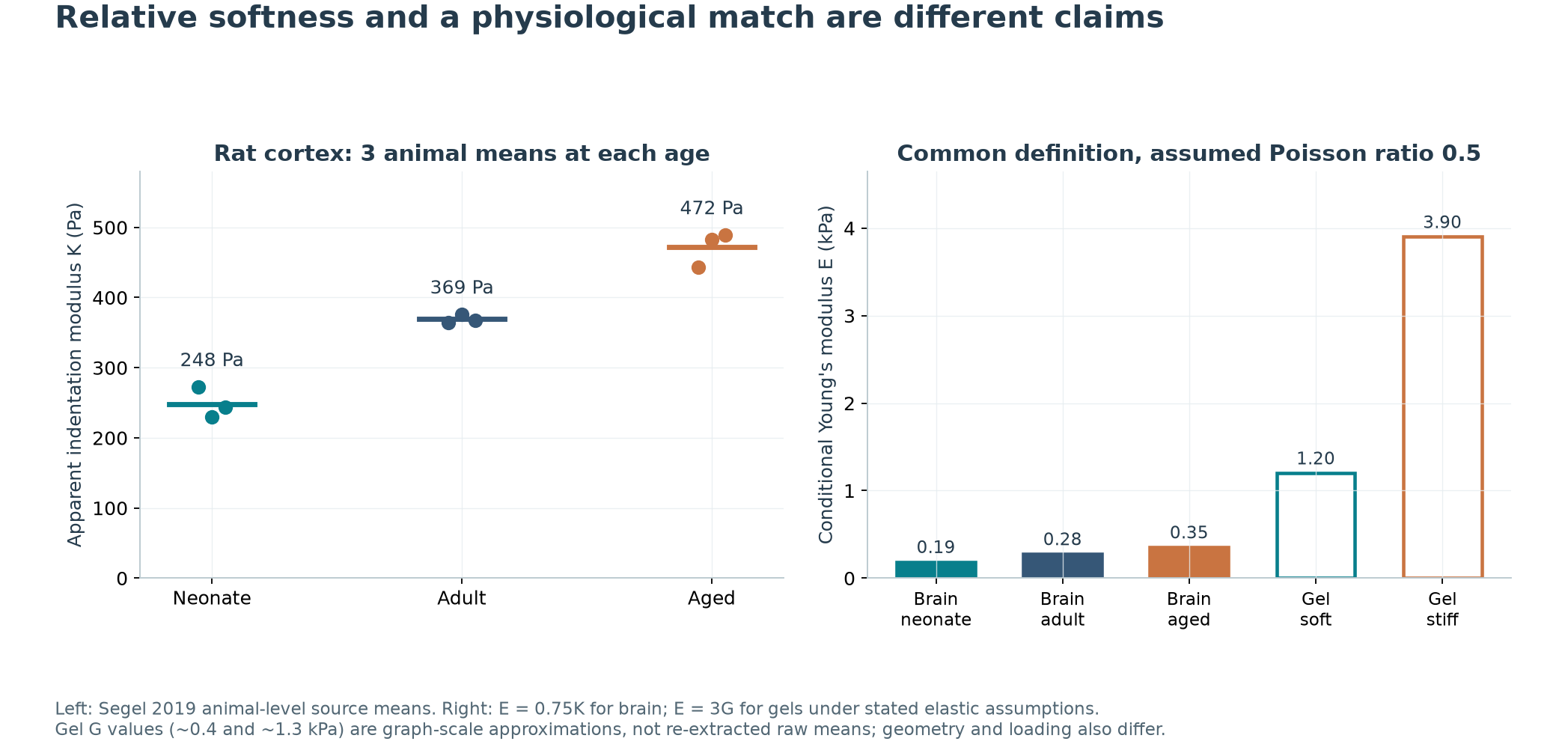
Converted to E under this assumption, the three tissue means are about 0.19, 0.28 and 0.35 kPa; the gels are about 1.2 and 3.9 kPa. Calling one gel relatively soft does not establish that it reproduces the absolute mechanics of young brain tissue.
This does not negate the observed gel effects. It limits attempts to translate culture results into a physiological threshold. Brain tissue is also viscoelastic, directional and structurally complex, and probe indentation differs from cellular traction in its loading conditions.
Scale matters in human research too. A 2015 study of 45 cognitively normal, amyloid-PET-negative participants aged 56–89 used magnetic resonance elastography and found lower readings at older ages in several, but not all, brain regions. Human brain mechanics study This dynamic, larger-scale, cross-sectional measurement differs from local indentation of rat slices in species, age window and measurement conditions. The results cannot be merged into a universal curve in which all brain tissue stiffens with age.
What changed in the animal source tables?
In 18-month-old mice, investigators used viral delivery of a gene-editing strategy to reduce OPC-associated PIEZO1 expression before inducing a focal demyelinating lesion. Targeting was partial and varied between cells; it should not be understood as an identical, complete knockout in every OPC. Each of three primary source-table outcomes has three animal records per condition. Source 1
Our animal-level calculations give:
- The source-defined EdU/GFP/OLIG2 labeling fraction: about 13.7% in controls and 33.2% after targeting, a difference of 19.4 percentage points.
- Density of mature cells jointly labeled CC1 and OLIG2: about 64.0 versus 121.8 cells/mm², a difference of 57.8 cells/mm².
- FluoroMyelin-positive area fraction: about 8.2% versus 29.1%, a difference of 21.0 percentage points.
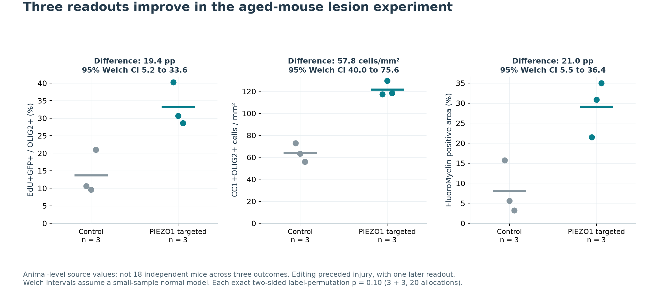
Approximate 95% Welch intervals are 5.2–33.6 percentage points, 40.0–75.6 cells/mm² and 5.5–36.4 percentage points, respectively. After Holm adjustment across these three exploratory comparisons, p values under that parametric model remain below 0.05.
With three animals per group, however, distributional assumptions cannot be checked adequately. In a separate sensitivity analysis, assuming group labels are exchangeable, a 3 + 3 design has 20 possible allocations. The exact two-sided permutation p value is 0.10 for all three outcomes. Such a small design encounters this resolution limit even when every treated value exceeds every control value.
Consistent observed directions and precise, generalizable effect estimates represent different strengths of conclusion. The tests rely on different assumptions; selecting one number cannot replace an assessment of the evidence. Nor do three outcomes constitute three independent animal replications: 18 measurements across the outcomes do not establish 18 independent mice.
More myelin staining can arise through different processes
Timing matters. The formal Figure 4 caption places viral delivery 14 days before injury and tissue collection 14 days after injury. More myelin staining at the later endpoint could therefore reflect increased formation, reduced continuing loss, a smaller initial injury, or some combination. Source 1
To illustrate, let M represent an idealized covered fraction, a a formation coefficient and b a loss coefficient:
dM/dt = a × (1 − M) − b × M
For fixed, nonnegative a and b with a positive sum, long-term coverage is a / (a + b). Different formation and loss rates can produce the same coverage. At a finite observation time, the initial injury also matters.
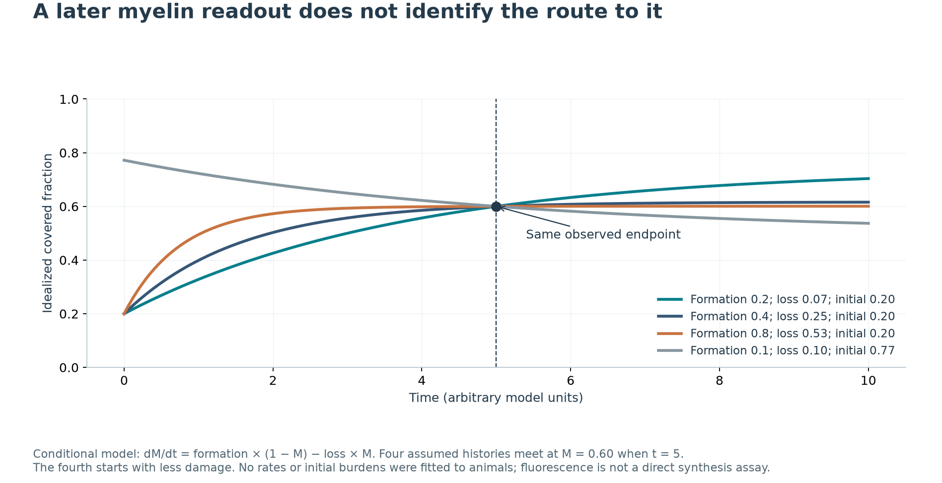
We constructed four assumed trajectories that reach 60% coverage at five model time units. Three begin at the same starting point with different formation and loss parameters. The fourth starts with less initial damage. All pass through the same observation.
These parameters were not fitted to mice, and model time is not measured in days. The model shows that one endpoint cannot uniquely identify the process; it does not establish which protective or repair mechanism actually operated in the experiment. FluoroMyelin staining is itself not a direct measurement of the rate of new myelin synthesis.
The original study also injected chABC or a cytoskeletal perturbation tool seven days after injury. Those experiments address a later intervention window. However, chABC changes matrix molecules, and cytoskeletal perturbation can affect multiple cells; their effects still need to be distinguished from changing stiffness alone.
Another study published online in 2019 reported reduced demyelination when the mechanosensitive-channel blocker GsMTx4 was given alongside the injury-inducing agent. We obtained the formal abstract and did not complete a full-text review. This co-administration design supports investigating protection; it cannot be rewritten as completed repair of an established aged lesion. Pharmacological study abstract
Molecular “youth” depends on the reference
The original study also used transcript-expression clustering to support a change in cellular state. We extracted 18 processed expression files from GEO, reconstructed abundances for 17,337 genes, and checked the aggregation for every sample using a separate R program. Public data
Twelve samples explicitly identify age and culture substrate: aged-soft, aged-stiff, neonatal-soft and neonatal-stiff, with three samples each. GEO annotations do not independently establish the detailed culture versus fresh-isolation identities of A1–3 and N1–3. We therefore exclude these six from the primary analysis and report a conditional reference analysis separately.
The primary analysis retains 15,017 genes with mean TPM of at least one under a rule set before the effect calculations. It compares equally weighted log2(TPM + 1) values. The distance describes how different two group-average expression profiles are. It is not a validated aging clock, and normalized abundances were not treated as raw counts for differential testing.
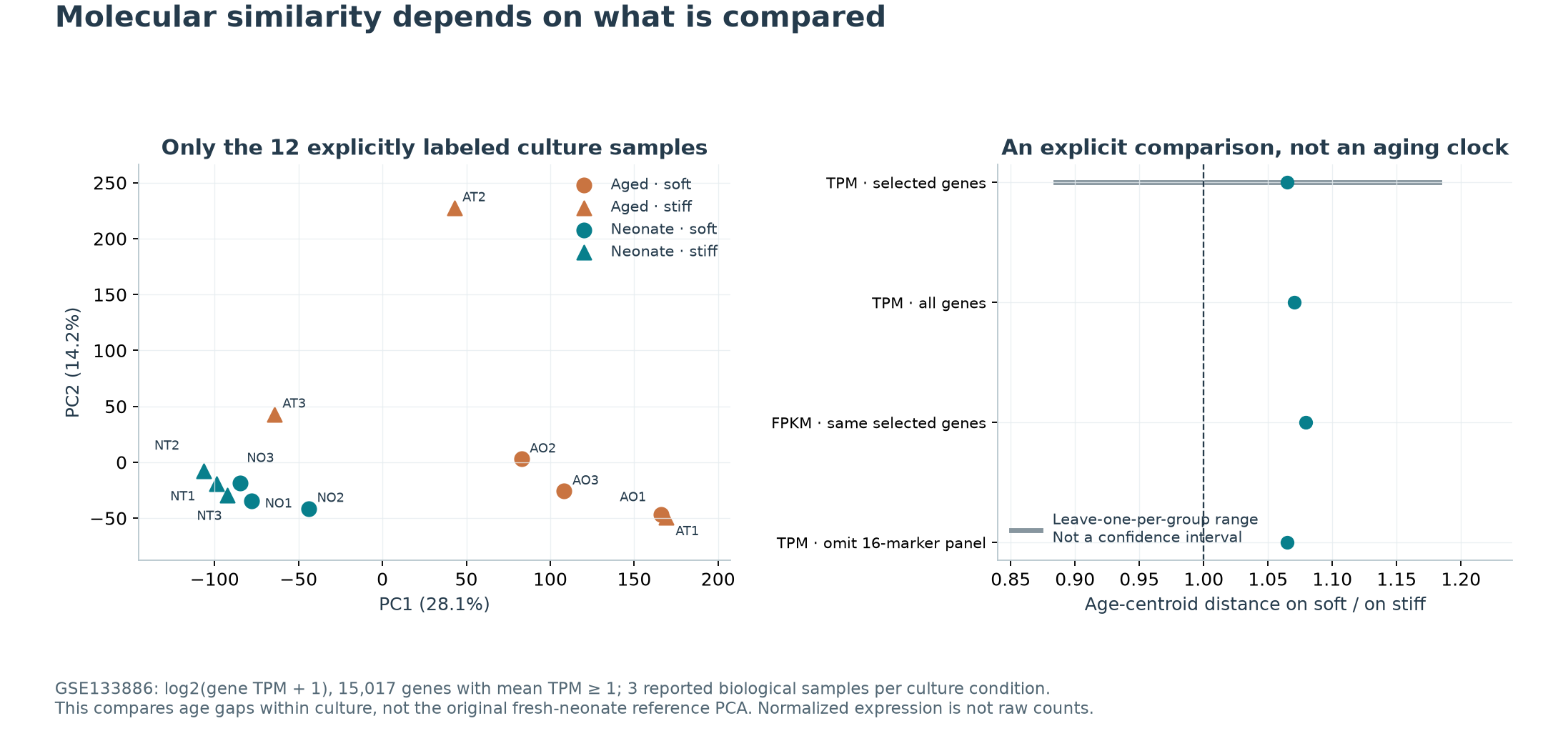
Two related but distinct questions give the following results:
| Comparison | Our result | Interpretation |
|---|---|---|
| Within a shared culture environment, is the aged–neonatal expression gap smaller on soft material? | Soft/stiff age-distance ratio about 1.065; all genes, FPKM or exclusion of the predeclared 16 cell-cycle markers gives about 1.065–1.079. Leaving one sample out per group gives a range of about 0.885–1.183 | This definition does not show a stable narrowing. The leave-one-out range is not a confidence interval, and this does not establish that soft culture makes cells older |
| Assuming from the naming that N1–3 are fresh-neonatal references, are aged-soft cultures closer to them than aged-stiff cultures? | Distance ratio about 1.068 in the main setting and 1.069–1.101 in the other settings; conditional leave-one-out range about 0.920–1.145 | Reference identity and processing annotations remain incomplete. This is an additional conditional calculation, not an exact reproduction of the authors' PCA |
Neither distance comparison directly measures cellular age. A gap within shared culture can narrow because young cells change, or persist even when function improves in both groups. Proximity to a reference also does not guarantee recovery of every function. We retain the positive culture and animal functional readouts while declining to turn a clustering plot into proof of complete rejuvenation.
Cell composition, culture selection, proliferative state, batch and undocumented donor pairing may affect expression. Removing 16 markers does not remove all cell-cycle effects. Determining whether the same aged cells recover durably requires clearer sample identities, cell tracking, functional measurements and follow-up after the environmental change is withdrawn.
Softer is not better under every condition
An earlier primary study in 2012 used neonatal rat-derived cells, a different substrate attachment chemistry and a broader stiffness range. Survival, proliferation and migration were favored at intermediate stiffness, while differentiation could increase with greater stiffness. Primary study under different culture conditions
Age, ligands, culture procedures, observation times and some measurement definitions differ from the 2019 study. It is not appropriate to declare one entire paper correct and the other incorrect from that comparison. Both support OPC responsiveness to mechanics. They do not establish a rule that progressively softening any environment improves every function.
Chemical signals also matter. Another 2019 study cultured OPCs on membrane and matrix material left by microglia and found that cell age, TGFβ treatment and related signals altered differentiation phenotypes. Microglial environment study The authors acknowledge that small numbers of contaminating cells could not be excluded as the source of some astrocyte phenotypes. Markers alone cannot establish that all such cells changed lineage. This study did not independently manipulate stiffness; it illustrates that replacing an environment can change several kinds of information.
Human material requires a separate interpretation. A 2022 study examined PIEZO1 in human brain samples and studied migration and MTT readouts in the MO3.13 cell line. Migration or metabolic-activity measurements in a cell line are not myelin repair in older patients. The human expression comparison also cannot establish that channel inhibition would benefit patients. Human tissue and cell-line study
Active cells still need to survive and complete repair
Driving progenitors toward differentiation and keeping newly formed mature cells alive are distinct stages. A simple model separates them. Let O be the mature-cell population, q the rate of entry into that pool and δ its death-rate constant. Let s represent the contribution of mature cells to coverage formation and λ the coverage-loss constant:
dO/dt = q − δO; dM/dt = s × O × (1 − M) − λ × M
With constant nonnegative parameters and positive δ, the long-term mature-cell population is q/δ. If formation or loss remains positive, corresponding coverage is s × q / (s × q + λ × δ). Increasing q may still leave poor coverage if cell death or myelin loss also rises.
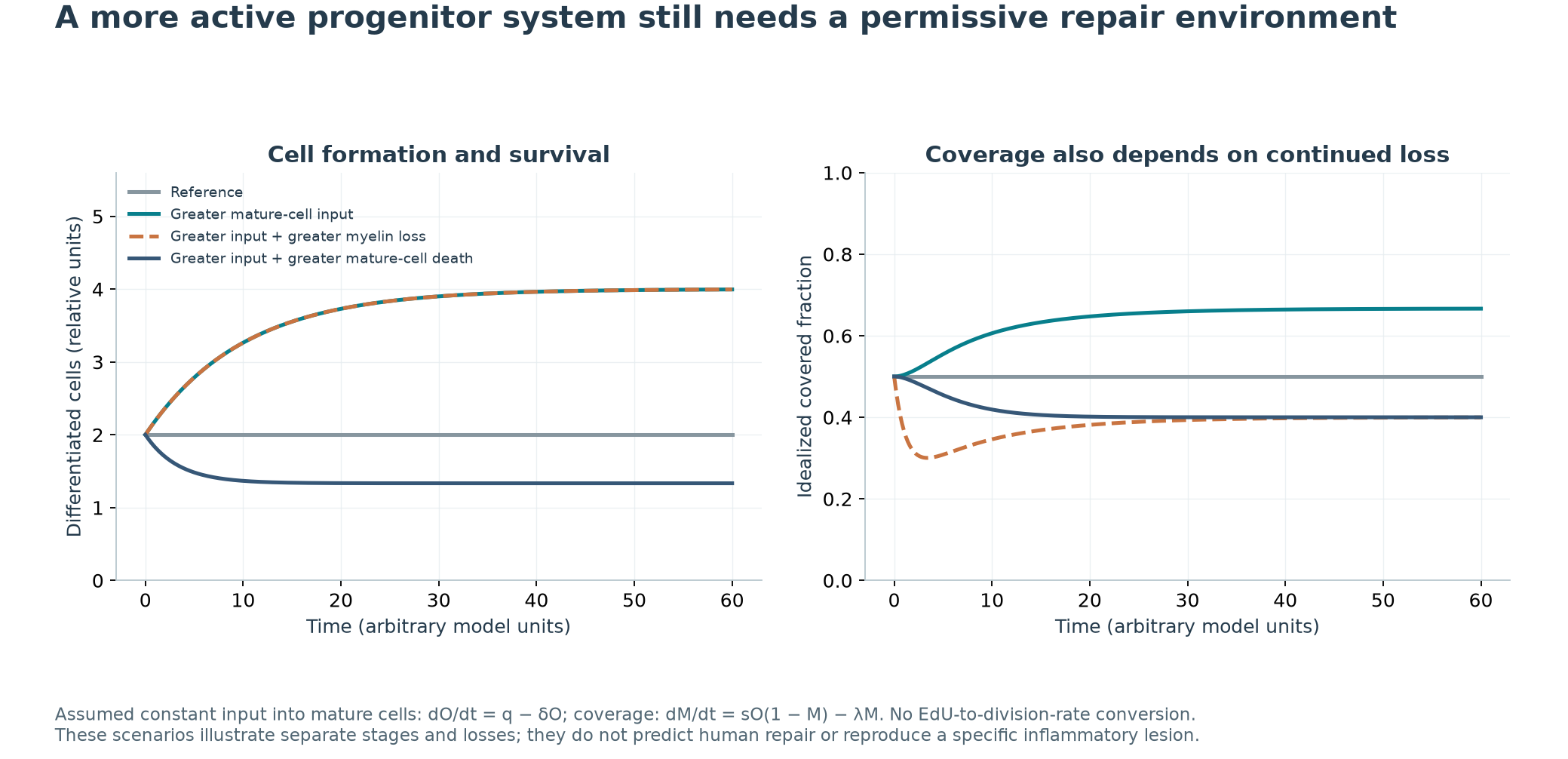
One assumed scenario doubles mature-cell input; another also increases myelin loss; a third also increases mature-cell death. The overlapping solid and orange dashed cell-number trajectories correspond to different coverage trajectories: equal cell numbers can accompany different final coverage. Arbitrary parameters illustrate relationships between stages. A single EdU-positive fraction was not converted into a division rate, and no inflammatory lesion was fitted.
The formal abstract of a 2026 immune-mediated demyelination study reports that OPCs in young and middle-aged animals can both respond to injury, while loss of differentiating cells and the inflammatory environment remain important. Recent study abstract This is neither a replication of the stiffness experiment nor evidence from very old humans. It supports distinguishing recruitment, differentiation, mature-cell survival and myelin maintenance.
Source discrepancies constrain specific conclusions
Several issues remain unresolved. The formal Figure 3 transplantation caption says four animals per group, but the available source table has three rows. The second Figure 1 transplantation block repeats a proliferation label; we cannot silently reassign it to differentiation. Two developmental cell-density blocks duplicate the same values and cannot independently verify two outcomes. The 563 indentation or cell-position measurements without animal identifiers were treated descriptively, not as 563 animals for significance testing.
One version difference was resolved: the early author-manuscript Figure 1 caption says three animals, whereas the current formal caption says four, matching the source table. The formal 2019 correction concerns an author name and funding; it does not establish that all the above source issues were corrected. Correction notice
There is also a discrepancy between random allocation in the author-manuscript Methods and no randomization in the publisher's reporting summary. We cannot certify allocation from that record. Some protein validation for the genetic intervention used transfected fibroblasts, so it should not be described as directly measuring an equivalent protein reduction in aged OPCs.
These issues do not erase every experiment's value. They affect the strength of inference a particular result can support.
What does “restoring regeneration” mean here?
The better-supported answer is that age-related declines in OPC activity include a component responsive to the environment and mechanical sensing. These experiments have not validated recovery of human neurological function, lasting rejuvenation or lifespan extension.
A more complete regeneration claim would connect initial injury comparability, cell survival and expansion, differentiation, new myelin formation, appropriate structure, improved conduction and behavior, and persistence after intervention withdrawal. Matrix chemistry, mechanics and continuing injury also need to be separated experimentally.
This connects the questions in the preceding two articles: clearance can affect damage accumulation, and damage can concentrate in a few locations. When repair starts, newly formed or reactivated cells still encounter the existing environment. Recovery requires these stages to work together. Softer material, more cells or greater staining each answers only part of the question.
Data and reproduction
Download the animal and expression extracts, statistical analyses, mechanism models and five-figure reproduction package. The 95 animal-level outcome records include reused cohorts and retained ambiguous data excluded from inference; they are not 95 independent animals.
We independently checked 449 XLSX numeric cells using XML, checked 12 animal values from the older XLS format against the supplementary PDF, and reaggregated all 18 GTFs in R to compare 624,132 gene-level TPM/FPKM values. Key statistics, expression distances and models were also checked using independent implementations. Raw sequencing, image segmentation, all in-vitro replicate data and human clinical effects were not revalidated. Actual reading scope, same-author review and remaining limits are recorded below.
Scope & limitations
- Primary evidence concerns neonatal/aged rodents and small lesion experiments; three animals per group cannot adequately test distributional assumptions, and three outcomes are not independent replications.
- Missing Figure 3 rows, duplicate labels/ED10 densities, some cached-summary ranges and allocation descriptions remain unresolved; source values were not repaired to match expectations.
- The 563 points lack animal identities and are descriptive. The 95 outcome records are not 95 unique animals; regional fractions lack denominators for whole-brain purity.
- Primary RNA analysis uses 12 cultures. Fresh A/N reference identity is unverified; no raw-count, paired-donor or longitudinal age prediction is made. The later reference calculation is explicitly conditional.
- Gel moduli are graph-scale approximations. K/E/G conversions do not remove geometry, loading, viscoelasticity or species differences.
- PIEZO1 targeting is partial and precedes injury. Myelin area does not separate initial injury, formation and loss; these data do not validate conduction, behavior, lasting rejuvenation or human efficacy.
- Mechanistic parameters are assumed. Constant nonnegative rates and positive denominators are equilibrium conditions; EdU labeling is not inverted to division rates.
- Claims from the 2019 pharmacology and 2026 inflammation studies are abstract-bounded. Full methods/safety/disclosures and all in-vitro replicate data were not verified.
- Independent source checks cover 449 XLSX cells, 12 XLS animal values and 624,132 RNA aggregates. Pooled XLS points lack a second BIFF-parser check; raw sequencing/imaging were not reprocessed.
Sources
- Segel et al. Niche stiffness underlies the ageing of central nervous system progenitor cells. Nature (2019)
primary animal and cell study with source data · Source version: 2019; doi:10.1038/s41586-019-1484-9; author manuscript and current publisher materials acquired 2026-09-21
Reading scope
Relevant sections
Read all 11 main results/discussion paragraphs, relevant methods, listed captions, supplementary Notes/Tables 5–11 and design reporting; not every reagent list exhaustively. Extracted seven workbooks: 95 animal-level outcome records, 718 numbers and 563 points lacking animal IDs. Records are not unique animals. Current Figure 1 N=4 matches data; Figure 3 N=4 versus three rows, duplicate labels/ED10 and allocation descriptions remain unresolved. Gel G approximately 400/1,300 Pa comes from visual graph scale, not raw means; the publisher image is not redistributed. Targeting is partial; MEF protein validation is not aged-OPC protein evidence. Funding and no-competing-interest statement read.
- Main results/discussion; isolation, culture, gels, AFM, RNA, lesion and CRISPR Methods
- Main Figures 1–4; Extended Data 2, 4, 5, 8, 9 and 10; current publisher Figures 1, 3 and 4
- Supplementary Notes, Tables 5–11, Reporting Summary; source workbooks MOESM3–9
- Segel et al. Author Correction: Niche stiffness underlies the ageing of central nervous system progenitor cells. Nature (2019)
author correction · Source version: 2019; doi:10.1038/s41586-019-1552-1
Reading scope
Relevant sections
Read the complete correction notice: author name and Royal Society funding. It does not certify correction of numerical, sample-count, label or randomization issues.
- Correction notice: author name and Royal Society funding
- NCBI GEO. GSE133886: OPC gene expression under age and substrate conditions
public processed expression dataset · Source version: GSE133886; last series update 2025-07-22; acquired 2026-09-21
Reading scope
Relevant sections
Parsed all 18 GTFs into 17,337 genes without missing-value zero imputation; primary analysis uses 12 explicitly labeled cultures. A/N identities are conditional from naming; generic GEO culture protocols do not independently establish fresh isolation. TPM/FPKM are not raw counts. Separate R parsing checked 624,132 gene aggregates against the original archive. Raw reads/alignment/quantification were not rerun; donor pairing is unverified.
- Series and all 18 sample annotations; SOFT family metadata
- All 18 StringTie GTF transcript records; RN6 reference annotation; gene aggregation
- Jagielska et al. Mechanical Environment Modulates Biological Properties of Oligodendrocyte Progenitor Cells. Stem Cells and Development (2012)
primary cell culture study · Source version: 2012; doi:10.1089/scd.2012.0189
Reading scope
Relevant sections
Read abstract, culture/PDL-gel/measurement methods, relevant results/discussion and disclosures. Neonatal rat mixed-culture origin; E approximately 0.1–70 kPa. Survival/proliferation/migration and differentiation respond differently; no universal softest optimum. No replicate-data reanalysis or claim of matched-age/ligand replication of 2019.
- Abstract; relevant OPC/gel/measurement methods, results and discussion
- Acknowledgments and Author Disclosure Statement
- Arani et al. Measuring the effects of aging and sex on regional brain stiffness with MR elastography in healthy older adults. NeuroImage (2015)
primary human imaging study · Source version: 2015; doi:10.1016/j.neuroimage.2015.02.016
Reading scope
Relevant sections
Read the listed sections: 45 cognitively normal, PET-negative participants aged 56–89; regional dynamic shear-related measurements with 60-Hz MRE. Cross-sectional regional differences are not annual within-person softening or direct equivalence to AFM K/culture gels. Individual images not reanalyzed; not every disclosure verified.
- Abstract; Methods 2.1–2.3; conclusion and relevant Discussion
- Acknowledgements
- Baror et al. Transforming growth factor-beta renders ageing microglia inhibitory to oligodendrocyte generation by CNS progenitors. Glia (2019)
primary cell and niche study · Source version: 2019; doi:10.1002/glia.23612
Reading scope
Relevant sections
Read abstract, ghost preparation and relevant differentiation/signaling results/discussion. Membrane/matrix chemistry is not an independent stiffness manipulation. Authors cannot fully exclude astrocyte contamination; NG2 is not exclusive to OPCs in inflammation. Markers do not prove all lineage conversion. Funding includes Medimmune; other commercial relationships not exhaustively verified.
- Abstract; isolation/ghost preparation; relevant Results 3.1, 3.4 and 3.6
- Discussion 4.1–4.2 and funding statement
- Velasco-Estevez et al. Mechanoreceptor Piezo1 Is Downregulated in Multiple Sclerosis Brain and Is Involved in the Maturation and Migration of Oligodendrocytes. Frontiers in Cellular Neuroscience (2022)
human tissue and cell-line study · Source version: 2022; doi:10.3389/fncel.2022.914985
Reading scope
Relevant sections
Read relevant human-tissue and MO3.13 methods/results. IHC has 5+5 donors; qPCR has 2 controls/5 MS donors. Cell-line MTT is not a direct division-rate assay; migration is not human myelin recovery. No case-data refit or therapeutic inference from expression direction. Funding/no-commercial-conflict statement read.
- Abstract; IHC, culture, migration and MTT methods; Results 3.3 and 3.5 with captions
- Funding and Conflict of Interest
- Velasco-Estevez et al. Inhibition of Piezo1 attenuates demyelination in the central nervous system. Glia (2019 online; 2020 issue)
primary study, abstract-only appraisal · Source version: Online 2019-10-09; doi:10.1002/glia.23722; PMID31596529
Reading scope
Abstract
Formal indexed abstract only; institutional PDF acquisition returned 403. GsMTx4/LPC co-administration addresses protection, not delayed repair or selective PIEZO1 genetic proof. Full methods, all safety endpoints and funding not verified.
- Formal indexed abstract only
- Fresenko et al. Aged oligodendrocyte progenitors retain the capacity to respond to an inflammatory insult. Experimental Neurology (2026)
primary animal study, abstract-bounded appraisal · Source version: Online 2026-06-10; October 2026 issue; doi:10.1016/j.expneurol.2026.115874; PMID42270005
Reading scope
Abstract
Claims rely on the locally preserved formal abstract. Publisher web excerpts of introduction and selected methods/discussion were also read, but no complete text or individual data obtained. Young/middle-aged autoimmune injury is not very-old human evidence or stiffness replication. Recruitment/differentiation and cell death remain distinct. Published online in June despite an October issue; funding not verified.
- Formal indexed abstract; publisher web excerpts of introduction and selected methods/discussion
Authorship & review
Author self-review · Codex (AI agent)
2026-09-21 · Same-author Codex review of neonatal/aged definitions, animal/outcome/point units, formal versus manuscript captions, source labels and denominators, partial PIEZO1 targeting and MEF validation. Treatment 14 days before injury is not delayed repair. Recomputed three n=3-per-group readouts with Welch/Holm and conditional exact permutations. Separated 12-culture expression geometry from the later conditional A/N-reference comparison; leave-one-out ranges are not CIs or age clocks. K/E/G conversions and two models have explicit nonnegative/equilibrium conditions, with no animal parameter fit. Checked 449 XLSX values, 12 XLS animal values and 624,132 RNA aggregates. All 576 R scalar comparisons across 293 checks passed; 30 outputs replayed byte-identically in a fresh directory and from the public ZIP. Compared both languages for values, negations, assumptions, five figures, two tables and English metadata. Revised publication-readiness self-score 9.10; not independent human professional review. Live publication checks are recorded separately.
Remaining limitations:
- Primary evidence concerns neonatal/aged rodents and small lesion experiments; three animals per group cannot adequately test distributional assumptions, and three outcomes are not independent replications.
- Missing Figure 3 rows, duplicate labels/ED10 densities, some cached-summary ranges and allocation descriptions remain unresolved; source values were not repaired to match expectations.
- The 563 points lack animal identities and are descriptive. The 95 outcome records are not 95 unique animals; regional fractions lack denominators for whole-brain purity.
- Primary RNA analysis uses 12 cultures. Fresh A/N reference identity is unverified; no raw-count, paired-donor or longitudinal age prediction is made. The later reference calculation is explicitly conditional.
- Gel moduli are graph-scale approximations. K/E/G conversions do not remove geometry, loading, viscoelasticity or species differences.
- PIEZO1 targeting is partial and precedes injury. Myelin area does not separate initial injury, formation and loss; these data do not validate conduction, behavior, lasting rejuvenation or human efficacy.
- Mechanistic parameters are assumed. Constant nonnegative rates and positive denominators are equilibrium conditions; EdU labeling is not inverted to division rates.
- Claims from the 2019 pharmacology and 2026 inflammation studies are abstract-bounded. Full methods/safety/disclosures and all in-vitro replicate data were not verified.
- Independent source checks cover 449 XLSX cells, 12 XLS animal values and 624,132 RNA aggregates. Pooled XLS points lack a second BIFF-parser check; raw sequencing/imaging were not reprocessed.
Editorial approval · Codex (AI agent)
2026-09-21 · Same-author Codex review of neonatal/aged definitions, animal/outcome/point units, formal versus manuscript captions, source labels and denominators, partial PIEZO1 targeting and MEF validation. Treatment 14 days before injury is not delayed repair. Recomputed three n=3-per-group readouts with Welch/Holm and conditional exact permutations. Separated 12-culture expression geometry from the later conditional A/N-reference comparison; leave-one-out ranges are not CIs or age clocks. K/E/G conversions and two models have explicit nonnegative/equilibrium conditions, with no animal parameter fit. Checked 449 XLSX values, 12 XLS animal values and 624,132 RNA aggregates. All 576 R scalar comparisons across 293 checks passed; 30 outputs replayed byte-identically in a fresh directory and from the public ZIP. Compared both languages for values, negations, assumptions, five figures, two tables and English metadata. Revised publication-readiness self-score 9.10; not independent human professional review. Live publication checks are recorded separately.
Translation check · Codex (AI agent)
· Same author compared both complete manuscripts and English metadata: age/species/timing, animal units, effects and intervals, conditional reference identity, mechanical definitions, model conditions, source discrepancies, figures and human limits. Not independent human language review.
Funding & interests
No product promotion or corporate commission was involved in this task. The same Codex agent performed research, self-review, translation and editing; this is not independent human professional review.
Funding of cited research
Segel reports ERC, MS Society, BBSRC, Adelson, Royal Society and Wellcome/MRC support and no competing interests. Jagielska reports HFSP support and no competing financial interests. Arani reports EB001981/AG16574 support. Baror reports MS Society, Medimmune, Adelson and Wellcome/MRC support. The 2022 human-tissue study reports EU Marie Sklodowska-Curie/Polish funding and no commercial/financial conflict. Other relationships and disclosures for the two abstract-bounded studies were not fully verified; absence is not assumed.