The answer we arrived at, first
"When could it help" currently has only a situational answer: the biologically credible, repeatedly demonstrated scenario in animals is a chronically high senescent-cell burden in a degenerative tissue context, treated with a targeted, intermittent clearance strategy. "When could it harm" has an equally concrete list: acute tissue repair, immune surveillance of premalignant cells, embryonic development, and the self-limiting phase of fibrosis — contexts in which senescent cells are part of the solution, not the lesion. And the human evidence underneath both judgments is still thin: the only systemic RCT in essentially healthy people missed its primary endpoint (no between-group difference in the bone-resorption marker CTx at 20 weeks; re-computed p=0.62); the most-cited "high-p16 subgroup benefit" survives our formal interaction test — which the paper never reported — only for radius BMD (p=0.004), a signal partly constructed from an unusual control-stratum trajectory and one that disappears under the alternative p16 assay definition. Most tellingly, within the subgroup where the skeletal signal appeared, none of 35 circulating SASP analytes changed differently between arms after treatment — bone moved where SASP did not. That is as consistent with "the effect came from dasatinib's direct bone pharmacology" or "small-subgroup noise" as with senescent-cell clearance. Calling senolytics "proven anti-aging drugs" overstates the evidence; calling them "a scam" ignores a real biological basis. The most defensible position today is: a mechanistically plausible class of candidate interventions whose value depends on which cells, which timing, and which tissue.
| What we did | What we got | What it does not answer |
|---|---|---|
| Downloaded the Farr 2024 Source data workbook (publisher supplement) with SHA256 archiving | Per-participant plotted values for every figure: bone markers, BMD, SASP, baseline characteristics, by arm × p16 stratum × time point | No per-participant continuous p16 values (only stratum membership) and no absolute baselines (only % change) — so continuous interaction and ANCOVA are impossible |
| Recomputed every printed cell of the paper | Primary endpoint, secondary endpoints, and subgroup medians with Wilcoxon p-values all reproduced (CTx 20 wk p=0.617 vs printed 0.611) | Agreement proves our reading and population definitions, not the underlying measurements; one cell is implementation-sensitive (T3 CTx 2 wk: ours p=0.053, printed 0.049 — it straddles 0.05) |
| Ran the formal interaction tests the paper did not report (permutation DiD) | Of 9 exploratory contrasts only radius BMD (p=0.004) and CTx at 4 wk (p=0.025) are nominally significant; the headline T3 P1NP 2 wk contrast has interaction p=0.26 — indistinguishable from the low-p16 stratum | Subgroups are post hoc, n≈10 per arm; our test is a difference-of-medians DiD, not a covariate-adjusted regression |
| Mechanism check: asked the trial's own SASP panel whether clearance happened | Baseline: only 4 of 35 analysable analytes nominally higher in T3 vs T1/T2, none survives FDR; within T3, zero of 35 analytes differed between arms on D0→D14 change | Circulating SASP is not tissue-level senescent-cell burden; a plasma panel may miss local clearance (the paper's panel lists 36; one analyte was not released with the source data) |
| Control-arm stratum trajectory check (regression-to-mean / prognosis) | Within the control arm, T3 radius-BMD trajectories were significantly worse than T1/T2 (−1.5% vs +2.3%, p=0.006) — part of the "benefit" is built from the control side | Cannot separate prognostic meaning of p16 from small-sample chance |
| Mapped the animal and human evidence landscape | Genetic clearance extended median lifespan 24–27% in naturally aged mice; D+Q extended post-treatment survival ~36%; one human target-engagement signal (adipose p16+ cells fell); UBX0101 phase 2 failed; four independent lines for protective senescence | Most human data are open-label pilots with n<20; the UBX0101 result exists only at company-announcement level |
Scope: a layered evidence assessment of when clearing senescent cells is biologically grounded, when there are reasons for concern, and how far the existing human randomized evidence actually reaches. This is evidence description and provides no medication, dosing, or intervention advice; figures such as dasatinib 100 mg and quercetin 1000 mg appear only as the trial's registered protocol facts. Every help/harm judgment carries a species and context qualifier and is not a personal medical inference.
Unpacking the claim first
The title hides a conflation worth separating. A "senescent cell" is not a single entity: by trigger there is damage-induced senescence (DNA damage, telomere attrition, oxidative stress), programmed developmental senescence (cells scheduled to senesce during embryogenesis), and therapy-induced senescence (after chemo/radiation); by location, bone, fat, lung, skin, liver, immune cells; by persistence, transient cells that complete a signaling task and are then cleared by the immune system versus chronic stragglers. The markers they share — p16, p21, SA-β-gal, the SASP, Lamin B1 loss — each circumscribe overlapping but non-identical populations, and no single universal assay can declare "this is a senescent cell." "Clearing senescent cells helps" always means some subpopulation, in some tissue, at some time.
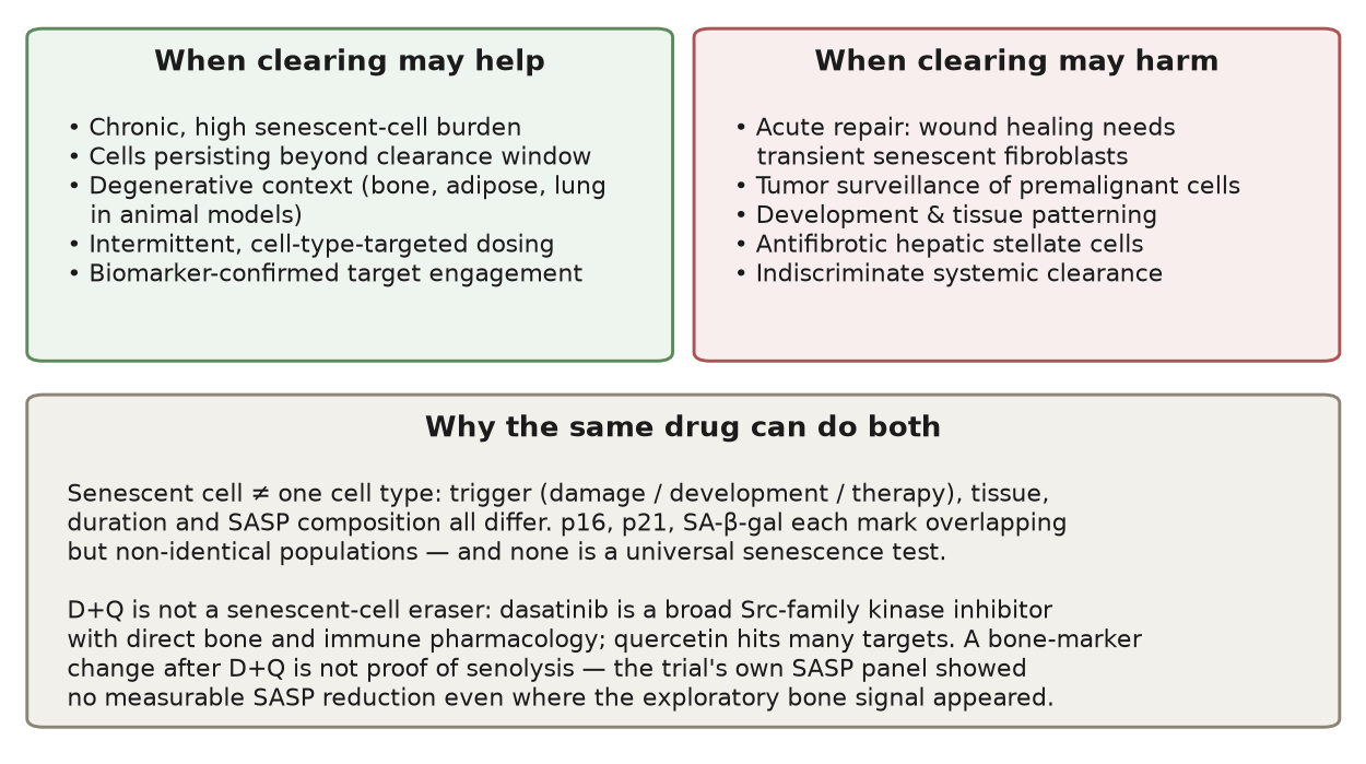
The clearing tools are not one blade either. Genetic tools (INK-ATTAC) can trigger apoptosis precisely in p16-expressing cells — but exist only in mice. Pharmacological senolytics grew out of senescent cells' anti-apoptotic dependencies (SCAPs): Zhu et al. 2015 showed that dasatinib and quercetin hit different SCAP nodes and kill different senescent cell types. Zhu 2015 But selectivity is relative: dasatinib began life as a leukemia drug and is a broad Src-family kinase inhibitor — and Src happens to be a kinase osteoclasts require to resorb bone, giving it a direct bone-pharmacology channel entirely unrelated to senolysis. Quercetin hits dozens of targets. Treating D+Q as a "senescent-cell eraser" is an oversimplification: most of what it does in the body is not senolysis. That point becomes an unavoidable competing explanation when we interpret the Farr trial.
Our analytic frame is therefore three questions that must be answered together: burden (are there really many cells worth clearing in this tissue), context (are those cells doing harm or doing a job right now), and attribution (did the observed effect come from clearance or from the drug's other pharmacology). The Farr trial happens to put all three on the table.
Human evidence: what one RCT can tell us
The trial and what it actually measured
Farr et al. 2024 is the highest-grade human evidence to date for systemic senolytic administration in essentially healthy older adults: a single-site, open-label, randomized, parallel-controlled phase 2 trial of 60 postmenopausal women comparing intermittent D+Q (registered protocol: dasatinib 100 mg × 2 days plus quercetin 1000 mg × 3 days per 28-day cycle, five cycles over 20 weeks) against untreated control. Farr 2024 Trial registry NCT04313634 The registry's actual enrollment of 74 reflects a third, fisetin arm that was discontinued and never entered the published analysis — so "74 vs 60" is protocol history, not a discrepancy.
Three design facts must stand before any number is read: open-label means adverse-event reporting and patient expectations are unblinded (reported events were mostly mild and concentrated in the D+Q arm — 77% of D+Q participants reported an adverse event vs 17% on control, with no serious adverse events); the primary endpoint CTx (bone resorption), secondary P1NP (bone formation), and BMD are bone-related surrogates — not lifespan, not broad healthspan, not a direct readout of "did people get younger"; and 20 weeks is short for a slow variable like aging. The trial answers "did D+Q change bone-metabolism markers over 20 weeks in postmenopausal women," not "does senolytic anti-aging work" — the latter needs a far larger evidence base.
Baseline was broadly balanced across arms (we recomputed the source-data Table 1 — age, weight, BMI, bone markers and site BMDs; the only nominal difference was serum calcium, 9.5 vs 9.3 mg/dL, p=0.027, a negligible magnitude). Attrition was asymmetric: 2 control participants terminated early, while the D+Q arm lost 3 — one withdrawn after QTc prolongation following dasatinib dosing (a known cardiac-conduction safety signal of the drug), one at week 9 for fatigue and nonspecific complaints, one after week 2 over COVID concerns. By week 20, markers were analysed on 28/28 available cases, BMD on 46–57 per site, and the T3 cells shrank from 11+10 to 10+8 — every denominator counts participants actually present, so there is no fiction of "60 completers."
Primary endpoint null; the secondary signal flickered out
The source-data workbook (publisher supplement MOESM3) gives per-participant plotted values for every figure. We first validated our inputs by recomputing every printed median, IQR and Wilcoxon p-value — primary, secondary, and subgroup cells all reproduced. [Figure 1]
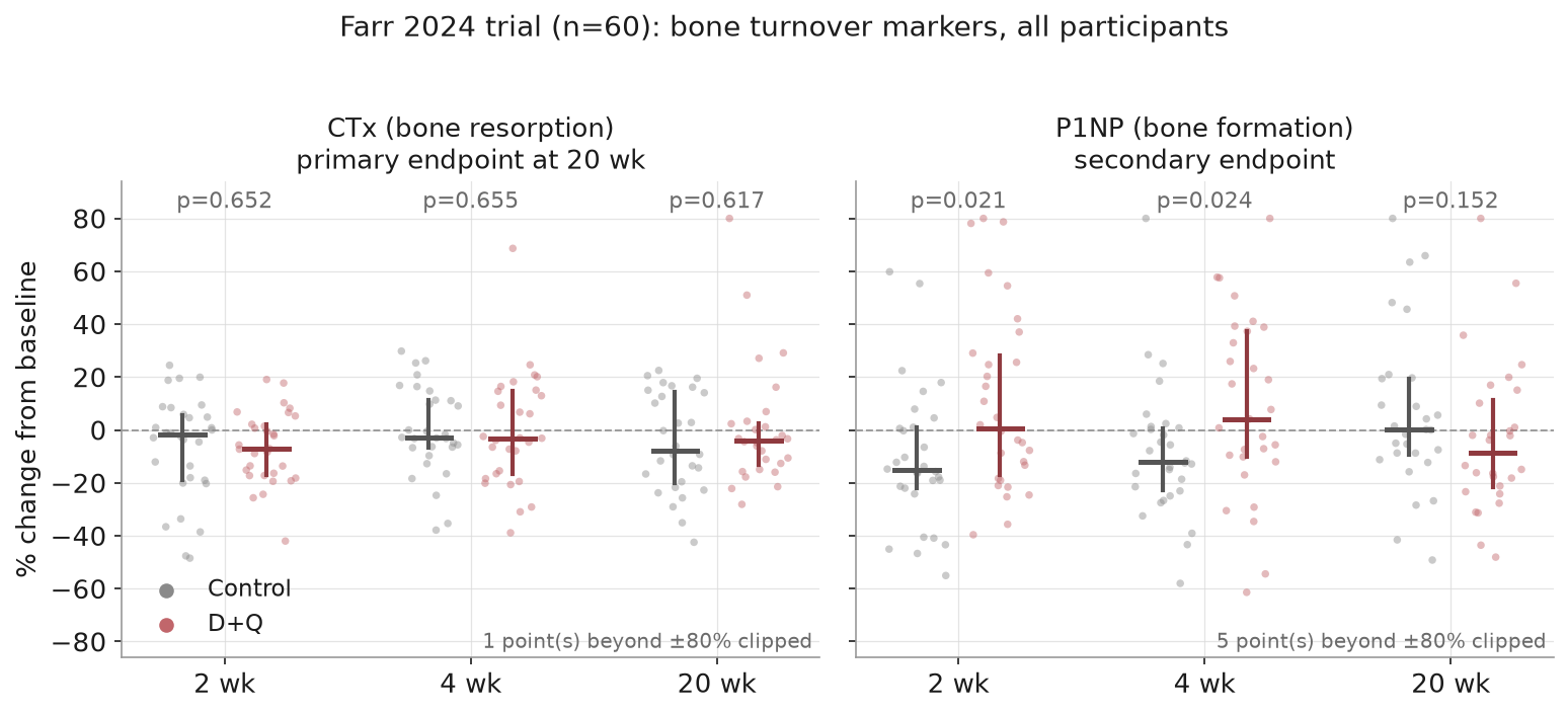
- Primary endpoint (CTx at 20 wk): null. Control median −7.7%, D+Q −4.1%, difference +3.6 points; re-computed p=0.617 (printed 0.611). The arms are indistinguishable on bone resorption at 20 weeks.
- Secondary (P1NP): a transient rise, then gone. +16.0 points relative to control at 2 weeks (re-computed p=0.021, printed 0.020), +16.2 at 4 weeks (p=0.024), then −8.7 at 20 weeks (p=0.15). The bone-formation marker twitched early and left nothing behind.
- BMD overall: no difference at any of three sites (radius, femoral neck, lumbar spine; n=55/57/46).
For a phase 2 trial with a null primary endpoint, the conventional story ends here. But the paper's real selling point — and the part most widely relayed — is its exploratory subgroup.
The high-p16 subgroup: the test the paper did not run
The authors split participants into tertiles by baseline T-cell p16 (variant-5 transcript) and called the top tertile (T3, ~21 women) the "high senescent-burden" stratum. Within T3 they reported: P1NP +34% relative to control at 2 weeks (p=0.035), CTx −11% at 2 weeks (p=0.049), and radius BMD +2.7% at 20 weeks (p=0.004) — concluding the skeletal response was "driven principally by women with a high senescent cell burden."
A statistical common-place must be stated first: "significant in subgroup A, not significant in subgroup B" is not evidence that A and B differ. Proving "driven by T3" requires a formal interaction test — asking whether the treatment-effect difference between strata is itself significant. The paper never reported one; we ran it on the source data: take the D+Q−control difference of medians within each stratum, then the difference of those differences (DiD), with a stratum-preserving permutation test (20,000 shuffles). [Figure 2]
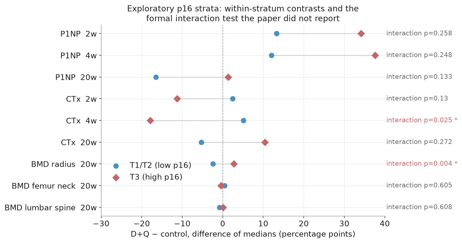
The result narrows "driven by T3" considerably:
- Of 9 exploratory contrasts, only 2 nominally pass interaction: radius BMD (DiD 5.1 points, p=0.004) and CTx at 4 weeks (p=0.025 — a time point the paper did not highlight).
- The most-quoted signal, T3 P1NP at 2 weeks, has interaction p=0.258 — although the within-T3 contrast is nominally significant (our re-computation p=0.038), its effect is statistically indistinguishable from the T1/T2 stratum's. "High-p16 participants benefit" does not stand on this marker.
- Applying multiplicity correction (Holm/BH) across the whole 18-cell exploratory family, only radius BMD survives (adjusted p=0.046); everything else disappears. Worth recording as calibration noise: one cell went nominally significant in the wrong stratum — T1/T2 P1NP at 20 weeks, −16.5% (p=0.042) — exactly the shape of randomness expected across many small cells.
Does the sole survivor — radius BMD — survive dissection?
Radius BMD is the only nominally significant interaction. One more layer of dissection shows what it is made of. [Figure 3]
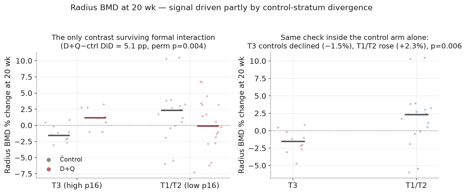
Within T3: D+Q median +1.2%, control −1.5%, a 2.7-point gap. But look inside the control arm alone — T3 controls ran a significantly worse trajectory than T1/T2 controls (−1.5% vs +2.3%, p=0.006). A substantial part of the "benefit" is not that D+Q-treated T3 participants did well (+1.2% is barely above zero and below the T1/T2 control median of +2.3%) but that T3 controls fell unusually. The two p16 strata differed in their 20-week natural course — which could be prognostic information carried by p16, a site-specific property of the radius (mostly cortical bone), or simply sampling noise in cells of 11 vs 17 women. The data cannot distinguish these. Either way, the signal is further discounted as evidence of treatment-effect modification: it fits "an unusual control-stratum trajectory" at least as well.
There is also assay sensitivity. The authors themselves ran a variant check: re-stratifying by p16 variant 1+5 (the routine clinical assay). Our re-computation shows the same cells under v1+5 — the strata themselves change first (T3 goes from 11+10 to 13+7, meaning a different set of women), effect sizes shrink, and every nominal significance vanishes (P1NP 2 wk goes from p=0.038 to 0.157). [Figure 4]
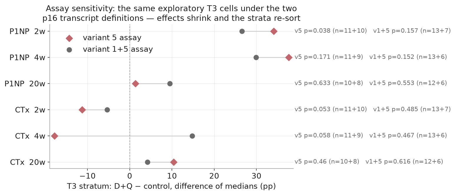
A signal that depends on which transcript defines "senescent burden" is a long way from established subgroup efficacy — it is hypothesis-generating material, nothing more, and the paper's own wording mostly stayed on that side of the line.
Mechanism check: where bone moved, did SASP move?
If the story is "clear senescent cells → SASP falls → bone metabolism improves," then circulating SASP analytes should fall precisely in the T3 stratum where the bone signal appeared. The source data let us test this directly: Extended Table 2 gives baseline SASP analytes by stratum; Extended Table 3 gives paired D0→D14 values for the 21 T3 participants (11 control, 10 D+Q). The paper describes a 36-analyte panel; the source data contain 35 analysable factors (numbering 1–36 skips item 9, which was not released). [Figure 5]
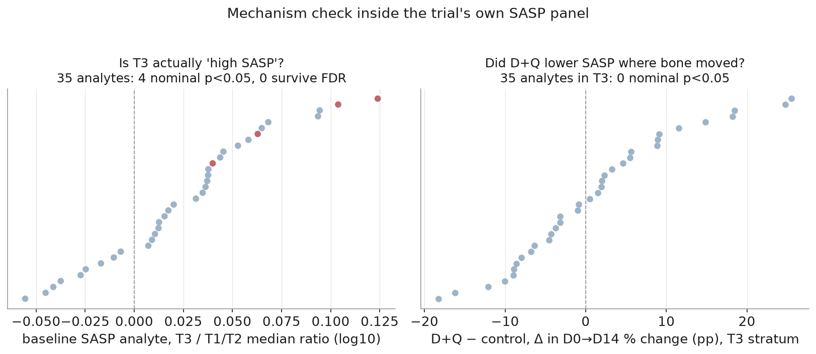
Two results are equally informative:
- "T3 is high-SASP" is itself weak. Median ratios for the 35 analytes mostly trend above T1/T2 (a consistent direction that may indicate a diffuse elevation), but only 4 analytes are nominally p<0.05 and none survives FDR correction. So the construct validity of "high T-cell p16 = high systemic senescent secretory burden" is thin — T3 is a proxy, not a clean marker of a high-SASP population.
- Within T3, zero of 35 analytes differed between arms on D0→D14 change (smallest p=0.149). The D+Q arm showed no measurable SASP reduction relative to control — where the bone signal appeared, the drug left no detectable trace of clearance. Worth recording: plasma SASP change was itself a registered secondary outcome of this trial — not an afterthought we dug up, but a pre-specified readout that came back empty.
An honest caveat must stand alongside: circulating SASP is not tissue-level senescent-cell burden, and a plasma panel could well miss local clearance; a paired test at n=21 has limited power. And an honest credit: the paper itself acknowledged that it "were not able to demonstrate effects of D+Q on circulating SASP markers" — what we add is the per-analyte quantification, the post-FDR construct-validity read, and connecting it back to the subgroup story: since the SASP panel was a registered secondary endpoint that came home empty, the "clearance → SASP drop → bone benefit" chain has no evidence inside this trial. But taken with the earlier findings — mostly-null interactions, a sole survivor partly built from control-arm divergence, assay-sensitive stratification — a simpler explanation surfaces: the observed bone-marker changes may not be senolysis at all, but dasatinib's direct bone pharmacology. Dasatinib inhibits Src-family kinases, and c-Src is required for osteoclast bone resorption — Src-deficient mice are osteopetrotic. An early CTx dip and a transient P1NP fluctuation are not inconsistent with "a Src inhibitor touched bone metabolism." The trial measured nothing that can separate the two channels (no tissue senescent-cell counts), so "clearance" and "pharmacology" cannot be distinguished in these data — which also means the result cannot be booked to senolysis's account.
The human evidence landscape: thin, with gaps everywhere
Putting Farr back into the human evidence chain makes the shape clear:
| Study | Design | n | What it measured | What it cannot say |
|---|---|---|---|---|
| Justice 2019, IPF | Open-label pilot | 14 | Intermittent D+Q feasible; functional signals | No control — cannot separate effect from natural fluctuation |
| Hickson 2019, diabetic kidney disease | Open-label | 9 | Adipose p16+/p21+/SA-β-gal+ cells down at 11 days — first human "target engagement" | n=9, uncontrolled, no clinical endpoint |
| Gonzales 2023, early Alzheimer's | Open-label | 5 | Dasatinib reached CSF (CNS penetration); quercetin undetected | Cognitive/imaging endpoints unchanged |
| Nambiar 2023, IPF | Single-blind placebo-controlled pilot | 12 | Feasibility/tolerability; sleep disturbance and anxiety clustered in D+Q arm | Not an efficacy trial, n=12 |
| Farr 2024, postmenopausal women | Open-label RCT | 60 | Everything re-computed in this article | Primary endpoint null; subgroups exploratory |
| UBX0101, knee osteoarthritis | Double-blind placebo-controlled phase 2 | 183 | No dose arm separated from placebo on WOMAC-A at 12 wk; program terminated (company-announcement level) | Intra-articular p53/MDM2 inhibitor — different mechanism and route from oral D+Q |
| UBX1325, diabetic macular edema/AMD | Intravitreal BCL-xL inhibitor | early | Visual-function signals with local dosing (contextual citation; primary text not retrieved) | Local lesion context — not systemic anti-aging evidence |
The chain's shape: feasibility is established (several pilots), target engagement has one preliminary demonstration (adipose biopsy), and no RCT has succeeded on a primary efficacy endpoint. This is not proof that senolytics do not work — it is "not yet shown to work," and the difference is the entire phase 2/3 pipeline.
Animal evidence: strong, but every "strong" carries a label
The numbers most often quoted for senolytics come from mice, and each is bound to a specific model:
- Baker 2011: INK-ATTAC clearance of p16+ cells in the BubR1 progeroid model delayed several age-related phenotypes. Concept proven — but a progeroid model is not natural aging. Baker 2011
- Baker 2016: in naturally aging wild-type mice, drug-induced INK-ATTAC clearance extended median lifespan 24–27% (27% mixed background, 24% C57BL/6, sexes combined), delayed tumorigenesis, and preserved organ function. The strongest animal case for "clearing p16+ cells helps" — but the vehicle is a transgenic switch no human can take, and p16 is only one proxy for senescence: what was cleared is "p16-expressing cells," not "all senescent cells." Baker 2016
- Xu 2018: transplanting senescent cells into young mice alone produced persistent physical dysfunction (evidence in the causal direction); intermittent D+Q in naturally aged mice extended post-treatment survival ~36% and reduced mortality hazard to 65% — often relayed as "36% longer life," actually a post-treatment survival-window metric, not a one-third extension of total lifespan. Xu 2018
- Khan 2025: temporal-lobe-epilepsy context — senescent glia were 5× elevated in human TLE tissue versus controls, and D+Q in the mouse model reduced senescent cells, rescued memory, and reduced seizures. Acquired senescence downstream of a defined lesion is precisely the context where senolysis is most plausible. Khan 2025
The honest strength of the animal evidence is: "in some models, in some tissues, for senescent cells defined by some marker, clearance improved function or lifespan" — it proves biological feasibility, not that any drug reproduces it in humans. From BubR1 progeria to natural aging, from a genetic switch to D+Q, each extrapolation had to be re-argued — let alone the leap to humans.
When it could harm: the other face of senescent cells
Clearing senescent cells can be a bad idea because the senescence program is part of the repair system; it becomes pathology mainly when cells overstay. Four independent lines of evidence:
- Embryonic development: programmed senescent cells perform structural remodeling during morphogenesis (Muñoz-Espín 2013 and related work) — scheduled "scaffolding cells" used and discarded. Disrupting them disrupts development.
- Wound healing: Demaria 2014 showed that senescent fibroblasts/endothelial cells appearing early in skin wounds accelerate closure by secreting PDGF-AA; removing them delayed healing, and PDGF-AA add-back rescued it. Demaria 2014 Acute SASP is a repair signal; chronic SASP is the inflammatory lesion — the same secretory phenotype, where the time scale decides good or bad.
- Tumor surveillance and antifibrosis: Krizhanovsky 2008 showed that activated hepatic stellate cells entering senescence after liver injury actually limit fibrosis — they stop proliferating, switch to secreting matrix-degrading enzymes, and actively summon NK cells to clear themselves; mice lacking senescence regulators got worse fibrosis. Krizhanovsky 2008 Senescence is also a proliferation brake on premalignant cells (Kang 2011 and related work): clearing the cells that stand watch could remove a cancer gate.
- The immune-clearance window: senescent cells are designed to exist briefly and then be removed by NK cells and macrophages. Part of why they accumulate with age is that immune clearance itself declines — which suggests two distinct intervention targets: reduce production, or restore clearance — not just periodic poisoning.
Put into one picture: the same dose may help in the "chronic high burden + degenerative context" cell of the grid and backfire in the "mid-repair, under-surveillance, or poorly selective" cells — and that is before adding the drugs' own toxicity (navitoclax's thrombocytopenia directly limited its clinical path; dasatinib carries pleural-effusion and myelosuppression risks in its oncology indication — not seen at intermittent low dose, but "not seen" is not "absent"). [Figure 6]
What this article can and cannot say
Can say: senescent-cell burden accumulates with age and participates in chronic inflammation via the SASP — supported in humans and animals; "clearance" strategies produced genuine lifespan and functional gains in mice; senescent cells have protective functions in development, wound repair, tumor surveillance and fibrosis self-limiting, each backed by independent primary evidence; the only systemic RCT in essentially healthy people to date missed its primary endpoint, and under our additional interaction tests, multiplicity correction, assay-sensitivity check and mechanism check only one exploratory cell barely survives — and that cell is partly constructed from an unusual control-arm trajectory; no human RCT has shown senolytics improving lifespan or broad healthspan outcomes.
Cannot say: senolytics are proven anti-aging drugs (no hard human endpoint supports that); senolytics are a scam (the animal evidence and target engagement are real — dismissing the whole field overreaches equally); the Farr trial "proved benefit in high-p16 participants" (interaction tests, assay sensitivity and the missing SASP response all argue otherwise); D+Q's bone-marker changes "prove clearance occurred" (the SASP panel did not move, and dasatinib's direct bone pharmacology remains an unexcluded competing explanation); or any dosing guidance — this is evidence description, and all dose figures are registered-protocol facts only.
Method boundaries: our re-computation rests on the publisher's Source-data plotted values — near-individual-level, but without per-participant continuous p16 or absolute baselines, so continuous interaction, ANCOVA, and individual responder tracking are impossible; subgroups are post hoc with ~10 participants per cell, where nominal p-values are inherently unstable; our interaction test is a difference-of-medians DiD with permutation, not a covariate-adjusted model; the circulating SASP panel cannot see local tissue clearance; safety data cover 20 weeks under an open label and cannot be extrapolated to long-term use.
So, the title's answer
When it might help: when senescent cells are chronically accumulated and the burden is measurable, when the tissue context is degenerative rather than mid-repair, when the clearing tool is selective for that cell type, and when dosing is intermittent rather than continuous — mouse evidence supports that combination, while humans have one preliminary target-engagement signal and a stack of feasibility studies. The scenario most likely to be established first is not "healthy older people take senolytics to prevent aging" but "indication-level trials in diseases with a defined senescent-cell-driven lesion" — IPF, osteoarthritis, retinal disease have all been tried, and their win-loss record is precisely the most honest part of the story.
When it might harm: whenever the cells are on the job — mid-wound-repair, premalignant surveillance, development, fibrosis self-limiting; or when burden is overestimated, markers misread, drug selectivity insufficient, so what gets cleared is functional tissue or what gets paid is drug toxicity without clearance in return. And the subtlest version: treating a "nominally significant post-hoc subgroup" as a prescribing criterion and giving the drug to people it was never shown to help.
What this field needs most right now is not more enthusiasm but three things: a biomarker that reliably measures senescent-cell burden in humans (a single T-cell p16 transcript clearly does not), controlled trials with clearance verification (tissue or a credible SASP response), and follow-up long enough to answer "how fast do they re-accumulate, and is repeated clearing safe." Until then, the most accurate description of senolytics is — a candidate strategy with a real biological basis, strong animal evidence, thin human evidence, and a "when it could harm" list that fills a page of its own.
Sources
- Farr 2024: intermittent senolytic therapy on bone metabolism in postmenopausal women — phase 2 RCT (full text + source data)
- Trial registry NCT04313634 (three-arm protocol history and endpoint definitions)
- Farr 2024 Source data workbook (per-participant plotted values, SHA256 archived)
- Zhu 2015: SCAP dependencies and D+Q cell-type selectivity
- Baker 2011: INK-ATTAC clearance of p16+ cells in a progeroid model
- Baker 2016: clearing p16+ cells in naturally aged mice extends median lifespan 24–27%
- Xu 2018: senescent-cell transplant causes dysfunction; D+Q +36% post-treatment survival
- Khan 2025: senescent glia in temporal lobe epilepsy and D+Q
- Demaria 2014: senescent cells accelerate wound healing via PDGF-AA
- Krizhanovsky 2008: senescent hepatic stellate cells limit fibrosis
- Hickson 2019: D+Q reduced adipose senescent-cell burden (n=9)
- Justice 2019: first-in-human D+Q pilot in IPF (n=14)
- Nambiar 2023: placebo-controlled IPF pilot (n=12)
- Gonzales 2023: SToMP-AD early-Alzheimer's pilot (n=5)
- UBX0101 phase 2 announcement: knee OA missed 12-week endpoint
- Muñoz-Espín, Kang, Ritschka: senescent-cell functions in development/surveillance (abstract level)
Scope & limitations
- The recomputation rests on the publisher's Source-data plotted values: near-individual-level but not raw case data; there are no per-participant continuous p16 values and no absolute baseline concentrations, so continuous interaction, ANCOVA and individual responder tracking are all impossible (c3, c5).
- The subgroups are post hoc with about 10 participants per cell, where nominal p-values are inherently unstable; the interaction test is a difference-of-medians DiD with permutation, not a covariate-adjusted model (c3).
- The control-stratum trajectory difference cannot be separated into 'prognostic information carried by p16' versus 'small-sample chance' (c5).
- A circulating SASP panel cannot see local tissue clearance; a null mechanism check does not exclude tissue-level clearance (c7).
- Open-label design: adverse-event reporting and patient expectations were unblinded; 20 weeks of follow-up is short for an ageing endpoint (c2, c12).
- Bone-metabolism markers and BMD are surrogate endpoints, not lifespan or broad healthspan readouts (c2).
- The UBX0101 phase-2 negative exists only at company-announcement level; UBX1325 and ongoing trials were not exhaustively searched (c11).
- The development and surveillance entries of the protective-function literature were read at abstract level (c10).
- Whether p16 variant 5 or variant 1+5 is the better assay is beyond what these data can decide; we report sensitivity, not an adjudication of the assays (c6).
- The self-review was performed by the same agent that authored the article; the new analyses (permutation tests, multiplicity correction) are seeded and their outputs archived, but implementation errors not covered by comparison against printed values remain possible.
Sources
- Farr JN et al. Effects of intermittent senolytic therapy on bone metabolism in postmenopausal women: a phase 2 RCT. Nat Med 2024
paper · Source version: PMC full text + Nature page snapshot, downloaded 2026-09-18
Reading scope
Full text
Full text read. Key facts verified: n=60 (30/30); primary endpoint CTx 20 wk P=0.611; P1NP 2w +16% P=0.020, 4w +16% P=0.024, 20w −9% P=0.149; T3 P1NP 2w +34% P=0.035, CTx 2w −11% P=0.049, radius BMD +2.7% P=0.004; subgroups exploratory and post hoc; variant 1+5 sensitivity did not preserve significance; no serious treatment-related AEs, minor AEs clustered in the D+Q arm under an open label.
- Results: primary/secondary endpoints, subgroups, safety
- Methods: open-label design, Wilcoxon convention, p16 assays (variant 5 vs 1+5)
- Figures 3/4/5 legends and n's
- ClinicalTrials.gov NCT04313634 registry record
trial_registry · Source version: API JSON snapshot 2026-09-18
Reading scope
Full text
Full registry record read. Confirms: actual enrollment 74 (including the discontinued fisetin arm — hence 60 in the published analysis); primary outcome CTx baseline→20 wk; secondary outcomes P1NP 2/4/20 wk, BMD 20 wk, SASP 2 wk; no masking; dosing schedule recorded as trial fact only (D 100 mg×2 d + Q 1000 mg×3 d per 28-day cycle, five cycles).
- design: randomized, parallel, no masking, phase 2
- arms: D+Q / fisetin / untreated control
- enrollment 74 vs published 60
- dosing schedule fields
- Farr 2024 Source data workbook (MOESM3 xlsx) and supplement PDFs MOESM1/2
dataset · Source version: Publisher supplements, downloaded 2026-09-18; SHA256 in the evidence manifest
Reading scope
Full text
All 20+ worksheets audited and parsed cell by cell: Fig 3a-f overall markers, Fig 4a-d stratified markers, Fig 5a-c stratified BMD, Extended Figs 4/5/6, Extended Table 2 (35 baseline SASP analytes by stratum), Extended Table 3 (paired D0→D14 SASP for 21 T3 participants), Table 1 (baseline characteristics). Confirmed absent: per-participant continuous p16 values and absolute baselines.
- data/41591_2024_3096_MOESM3_ESM.xlsx (per-participant plotted values for every figure)
- data/41591_2024_3096_MOESM1_ESM.pdf (visit schedule)
- data/41591_2024_3096_MOESM2_ESM.pdf (reporting checklist)
- Zhu Y et al. The Achilles' heel of senescent cells: from transcriptome to senolytic drugs. Aging Cell 2015
paper · Source version: PMC full text 2026-09-18
Reading scope
Full text
Full text read. Source for the SCAP concept and the pharmacological basis of the D+Q combination; also supports the point that the two drugs have different cell-type selectivity and each carries non-target pharmacology.
- SCAP dependencies argument
- D and Q cell-type selectivity
- Baker DJ et al. Clearance of p16Ink4a-positive senescent cells delays ageing-associated disorders. Nature 2011
paper · Source version: PMC full text 2026-09-18
Reading scope
Full text
Full text read. Proof-of-concept in a progeroid model; its boundary as a non-natural-aging model is stated.
- BubR1 progeroid model design
- phenotype-delay results
- Baker DJ et al. Naturally occurring p16Ink4a-positive cells shorten healthy lifespan. Nature 2016
paper · Source version: PMC full text 2026-09-18
Reading scope
Full text
Full text read. Strongest animal evidence; two qualifications written into the text — the vehicle is the INK-ATTAC transgenic switch, and what is cleared is p16-expressing cells, not all senescent cells.
- median lifespan +27% (mixed background) / +24% (C57BL/6)
- delayed tumorigenesis, preserved organ function
- non-tumour deaths 24-42%
- Xu M et al. Senolytics improve physical function and increase lifespan in old age. Nat Med 2018
paper · Source version: PMC full text 2026-09-18
Reading scope
Full text
Full text read. Two distinctions written into the text: the transplant experiment shows the causal direction, and +36% is a post-treatment survival-window metric, not a one-third extension of total lifespan.
- transplant model establishing the causal direction
- post-treatment survival +36%, mortality hazard 65%
- Khan T et al. Senescent cell clearance ameliorates temporal lobe epilepsy and associated spatial memory deficits in mice. Ann Neurol 2025
paper · Source version: PMC full text 2026-09-18
Reading scope
Full text
Full text archived; abstract and key results sentences verified. Used as an example of acquired senescence downstream of a defined lesion — not as human efficacy evidence.
- abstract/results: five-fold senescent glia in human TLE; D+Q reduces seizures and improves memory in the mouse model
- Demaria M et al. An essential role for senescent cells in optimal wound healing through secretion of PDGF-AA. Dev Cell 2014
paper · Source version: PMC full text 2026-09-18
Reading scope
Full text
Full text read. Primary evidence that acute SASP is a repair signal — load-bearing for the 'when it could harm' list.
- senescent fibroblasts/endothelial cells accelerate healing
- clearance delays healing; PDGF-AA add-back rescues
- Krizhanovsky V et al. Senescence of activated stellate cells limits liver fibrosis. Cell 2008
paper · Source version: PubMed abstract (efetch); PMCID PMC3073300 located
Reading scope
Abstract
Abstract-level read (an initial fetch pulled an unrelated SIRT1 paper; corrected to PMID 18724938). Load-bearing for the antifibrotic entry on the 'harm' list.
- abstract: senescent stellate cells limit fibrosis; NK clearance promotes resolution
- Hickson LJ et al. Senolytics decrease senescent cells in humans (DKD pilot). EBioMedicine 2019
paper · Source version: PMC full text 2026-09-18
Reading scope
Full text
Full text read. First human target-engagement evidence; boundaries stated — open label, n=9, no clinical endpoint.
- n=9 open-label
- adipose p16+/p21+/SA-beta-gal+ cells lower at 11 days
- Justice JN et al. Senolytics in IPF: first-in-human open-label pilot. EBioMedicine 2019
paper · Source version: PMC full text 2026-09-18
Reading scope
Full text
Full text read. First human D+Q administration; uncontrolled, functional readouts are signal-level.
- n=14 open-label pilot
- feasibility and functional signals
- Nambiar AM et al. D+Q in IPF: phase I single-blind placebo-controlled pilot. EBioMedicine 2023
paper · Source version: PubMed abstract (efetch); PMCID PMC10006434 located
Reading scope
Abstract
Abstract-level read. Better-controlled design but feasibility endpoints; the D+Q-arm AE pattern (sleep disturbance, anxiety) is recorded.
- n=12 placebo-controlled pilot
- sleep disturbance/anxiety clustered in the D+Q arm
- Gonzales MM et al. SToMP-AD: first senolytic trial in early Alzheimer's
paper · Source version: PubMed abstract (efetch)
Reading scope
Abstract
Abstract-level read. CNS penetration shown for dasatinib; no efficacy-endpoint change; cited only as one layer of the landscape.
- n=5 early-AD open-label
- dasatinib CSF penetration; quercetin undetected
- UNITY Biotechnology UBX0101 phase 2 announcement (GlobeNewswire, 2020-08-17)
press_release · Source version: Company announcement, web-retrieved 2026-09-18
Reading scope
Bibliographic record only
Announcement-level evidence: the phase 2 primary-endpoint miss and program termination are verifiable facts; numerical details not peer-reviewed and labelled accordingly.
- n=183 double-blind placebo-controlled
- no dose arm separated from placebo on 12-week WOMAC-A; programme terminated
- Muñoz-Espín 2013, Kang 2011, Ritschka 2017 — protective-function literature (abstracts)
paper_collection · Source version: PubMed abstract set 2026-09-18
Reading scope
Abstract
Abstract-level read. Backs the development and surveillance entries on the 'when it could harm' list; primary sources but not read to full-text depth.
- programmed senescence in development
- senescence surveillance of premalignant cells
Authorship & review
Author self-review · Devin (AI agent)
2026-09-18 · The author's own focused review of the whole article, performed by the same agent: agreement between the main question and scope; actual reading depth of load-bearing sources (Farr full text + per-sheet source data, the registry, full texts of Baker x2/Zhu/Xu/Hickson/Justice/Demaria/Khan; abstract-level for Krizhanovsky/Nambiar/Gonzales/Munoz-Espin/Kang/Ritschka; announcement-level for UBX0101); whether the analysis plan was locked before use; input validation (27 printed cells recomputed); whether statistics match their interpretation (difference-of-medians DiD permutation, not a covariate-adjusted model; an 18-cell multiplicity family; nominal vs adjusted p kept apart); retention of unfavourable and null signals (null primary endpoint, unmoved SASP panel, a reverse nominal T1/T2 cell, AEs clustered in the D+Q arm, the QTc withdrawal); the competing-explanation structure (direct dasatinib bone pharmacology vs senolysis); layered evidence rather than a single compressed rating; translation consistency and overstated claims; safety boundary (no dosing advice). Method: every load-bearing number was checked cell-by-cell against computation outputs and the paper/registry text, both language versions were compared for parity and source attribution, then a 0-10 self-score; two revision rounds preceded the final rating.
Remaining limitations:
- This self-review was performed by the same agent that authored the article; it is not an independent, human, or professional review. The permutation implementation was validated against printed values but may still contain errors not covered by that comparison.
- The source data are plotted near-individual values: no per-participant continuous p16 or absolute baselines, so continuous interaction, ANCOVA and individual responder tracking are impossible — conclusions stay at group level.
- Whether the control-arm T3 trajectory anomaly is prognostic information or small-sample chance cannot be resolved by these data; the radius-BMD signal is only partially decomposable.
- A circulating SASP panel can miss local tissue clearance; the null mechanism check does not exclude tissue-level clearance.
- Several protective-function and translational references were read at abstract or announcement level; ongoing trials were not exhaustively searched.
Editorial approval · Devin (AI agent)
2026-09-18 · Editorial sign-off executed by the same agent that authored the article, explicitly in a different working role — not independent, human, or professional review. Checked against the publication-blocking list: (1) source identity and versions — Farr full text/source data/supplements and the registry fetched under correct identifiers with SHA256 archiving; load-bearing PMCIDs verified one by one (a wrong Krizhanovsky fetch was corrected; the Khan 2025 attribution was miswritten as Millar and fixed in self-review); (2) no conclusion reversed relative to sources — null primary endpoint, unmoved SASP panel, a reverse nominal T1/T2 cell, AE clustering in the D+Q arm and the QTc withdrawal are all retained; (3) denominators and statistical populations — per-endpoint available cases, T3 cells 11+10 shrinking to 10+8, BMD site n=55/57/46, and the 11-vs-17 control-stratum trajectory comparison are stated as in the source data; (4) no model assumption presented as fact — post-hoc subgroups, the difference-of-medians DiD permutation test, and the 18-cell multiplicity family are labelled as such, and the unresolvable control-arm anomaly is stated; (5) safety boundary — no dosing advice, dose figures are registered-protocol facts only; (6) public scope is evidence description, so professional review is not triggered. Covers the current English text.
Translation check · Devin (AI agent)
· The same agent wrote the English text from the same evidence record rather than translating sentence by sentence, then compared it against the Chinese section by section. Checked: identical statistics (p=0.617/0.611 primary endpoint, +16.0/+16.2/-8.7pp P1NP, interaction p=0.004/0.025/0.258, control-stratum -1.5% vs +2.3% p=0.006, Holm 0.046, v1+5 restratification 11+10->13+7, 35 analysable SASP analytes with the 36-item panel note, 77% vs 17% AE reporting, 3 vs 2 withdrawals incl. the QTc case, corrected Khan 2025 attribution); every qualification preserved in English (post hoc subgroups, difference-of-medians DiD, registered-secondary-endpoint SASP null, contextual citation downgrade for UBX1325, announcement level for UBX0101, no dosing advice); no English-only claim added or dropped.
Funding & interests
This library is an independent research project with no commercial interest: it sells no products and provides no medical services, and it has no relationship with the authors of the cited papers, the journals, the Mayo Clinic, Unity Biotechnology, or any drug manufacturer. The author is an AI agent that also performed the editorial sign-off; the sign-off states this identity explicitly and does not claim independent review.
Funding of cited research
No external funding, and no drug- or supplement-related interest was accepted. Third-party relationships the reader should know: the Farr trial was run by academic institutions including the Mayo Clinic, with which this library has no affiliation; Unity Biotechnology is a listed company, and the UBX0101 result is cited at company-announcement level.